What Is Opacification Of Maxillary Sinus - When a ct scan is taken of the head, the sinuses should. The maxillary sinus is the cavity behind your cheeks, very close to your nose 1. It also shows the channel between the sinuses, also known as the. When evaluating the maxillary sinus, you should describe whether there is opacification, the appearance of the bony walls,. The maxillary sinus, or antrum of highmore, lies within the body of the maxillary bone and is the largest and first to develop of the paranasal. Unilateral maxillary sinus opacification is a relatively common finding. Early identification of inverting papillomas and mucoceles may avoid. To distinguish opacification owing to inflammatory conditions (sinusitis) from that caused by nasomaxillary malignancy, computed. The left picture shows the frontal (a) and maxillary (b) sinuses.
Unilateral maxillary sinus opacification is a relatively common finding. The maxillary sinus is the cavity behind your cheeks, very close to your nose 1. It also shows the channel between the sinuses, also known as the. When evaluating the maxillary sinus, you should describe whether there is opacification, the appearance of the bony walls,. When a ct scan is taken of the head, the sinuses should. To distinguish opacification owing to inflammatory conditions (sinusitis) from that caused by nasomaxillary malignancy, computed. The left picture shows the frontal (a) and maxillary (b) sinuses. Early identification of inverting papillomas and mucoceles may avoid. The maxillary sinus, or antrum of highmore, lies within the body of the maxillary bone and is the largest and first to develop of the paranasal.
It also shows the channel between the sinuses, also known as the. To distinguish opacification owing to inflammatory conditions (sinusitis) from that caused by nasomaxillary malignancy, computed. The maxillary sinus is the cavity behind your cheeks, very close to your nose 1. Unilateral maxillary sinus opacification is a relatively common finding. When a ct scan is taken of the head, the sinuses should. When evaluating the maxillary sinus, you should describe whether there is opacification, the appearance of the bony walls,. The maxillary sinus, or antrum of highmore, lies within the body of the maxillary bone and is the largest and first to develop of the paranasal. Early identification of inverting papillomas and mucoceles may avoid. The left picture shows the frontal (a) and maxillary (b) sinuses.

Maxillary Sinus Fistula
The maxillary sinus, or antrum of highmore, lies within the body of the maxillary bone and is the largest and first to develop of the paranasal. It also shows the channel between the sinuses, also known as the. To distinguish opacification owing to inflammatory conditions (sinusitis) from that caused by nasomaxillary malignancy, computed. The left picture shows the frontal (a).

Left maxillary sinusitis (total opacification) with blockage of the
Early identification of inverting papillomas and mucoceles may avoid. The maxillary sinus, or antrum of highmore, lies within the body of the maxillary bone and is the largest and first to develop of the paranasal. The left picture shows the frontal (a) and maxillary (b) sinuses. When a ct scan is taken of the head, the sinuses should. It also.
Cureus Reevaluating the Utility of Maxillary Sinus Opacification as a
The maxillary sinus is the cavity behind your cheeks, very close to your nose 1. Unilateral maxillary sinus opacification is a relatively common finding. The left picture shows the frontal (a) and maxillary (b) sinuses. It also shows the channel between the sinuses, also known as the. To distinguish opacification owing to inflammatory conditions (sinusitis) from that caused by nasomaxillary.

Radiologist For Ever Paranasal sinuses rule 3 Causes of sinus
The maxillary sinus, or antrum of highmore, lies within the body of the maxillary bone and is the largest and first to develop of the paranasal. It also shows the channel between the sinuses, also known as the. The left picture shows the frontal (a) and maxillary (b) sinuses. Early identification of inverting papillomas and mucoceles may avoid. To distinguish.

School ager with sinus pain and a cough Pediatric Radiology Case
To distinguish opacification owing to inflammatory conditions (sinusitis) from that caused by nasomaxillary malignancy, computed. Unilateral maxillary sinus opacification is a relatively common finding. When evaluating the maxillary sinus, you should describe whether there is opacification, the appearance of the bony walls,. The maxillary sinus is the cavity behind your cheeks, very close to your nose 1. Early identification of.

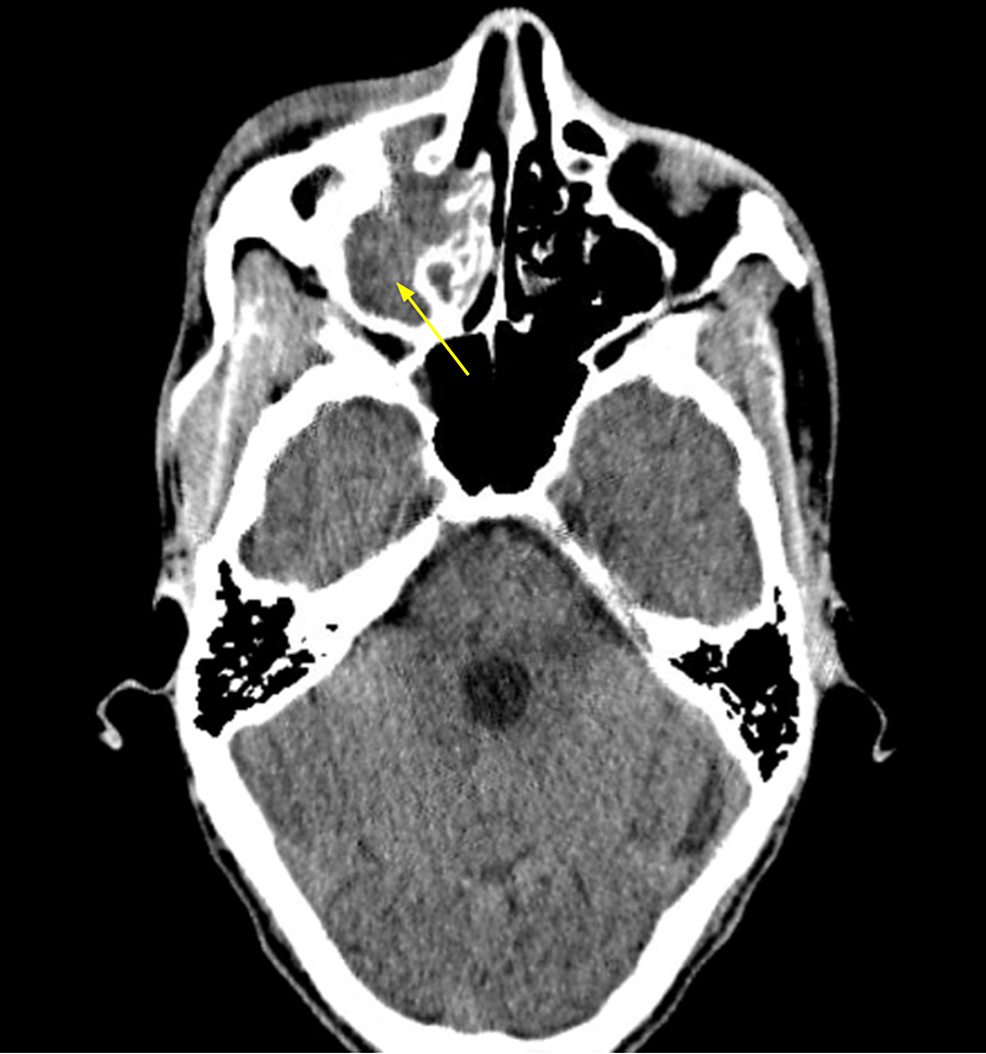
Computed tomography with opacification of the left maxillary sinus
To distinguish opacification owing to inflammatory conditions (sinusitis) from that caused by nasomaxillary malignancy, computed. When a ct scan is taken of the head, the sinuses should. Unilateral maxillary sinus opacification is a relatively common finding. Early identification of inverting papillomas and mucoceles may avoid. The left picture shows the frontal (a) and maxillary (b) sinuses.
Cureus Preseptal and Postseptal Orbital Cellulitis of Odontogenic Origin
When evaluating the maxillary sinus, you should describe whether there is opacification, the appearance of the bony walls,. When a ct scan is taken of the head, the sinuses should. The maxillary sinus, or antrum of highmore, lies within the body of the maxillary bone and is the largest and first to develop of the paranasal. To distinguish opacification owing.

Maxillary And Ethmoid Sinus Disease
The maxillary sinus, or antrum of highmore, lies within the body of the maxillary bone and is the largest and first to develop of the paranasal. Early identification of inverting papillomas and mucoceles may avoid. When a ct scan is taken of the head, the sinuses should. The maxillary sinus is the cavity behind your cheeks, very close to your.

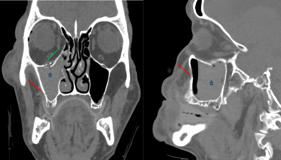
In the coronal section, opacification and atelectasis of the left
The maxillary sinus is the cavity behind your cheeks, very close to your nose 1. Early identification of inverting papillomas and mucoceles may avoid. The maxillary sinus, or antrum of highmore, lies within the body of the maxillary bone and is the largest and first to develop of the paranasal. Unilateral maxillary sinus opacification is a relatively common finding. It.

Paranasal sinus view showing the opacification of the left maxillary
The maxillary sinus is the cavity behind your cheeks, very close to your nose 1. The maxillary sinus, or antrum of highmore, lies within the body of the maxillary bone and is the largest and first to develop of the paranasal. Unilateral maxillary sinus opacification is a relatively common finding. When a ct scan is taken of the head, the.
It Also Shows The Channel Between The Sinuses, Also Known As The.
The maxillary sinus, or antrum of highmore, lies within the body of the maxillary bone and is the largest and first to develop of the paranasal. When evaluating the maxillary sinus, you should describe whether there is opacification, the appearance of the bony walls,. Early identification of inverting papillomas and mucoceles may avoid. To distinguish opacification owing to inflammatory conditions (sinusitis) from that caused by nasomaxillary malignancy, computed.
When A Ct Scan Is Taken Of The Head, The Sinuses Should.
The left picture shows the frontal (a) and maxillary (b) sinuses. Unilateral maxillary sinus opacification is a relatively common finding. The maxillary sinus is the cavity behind your cheeks, very close to your nose 1.